- 2014
Real-time 3D image reconstruction guidance in liver resection surgery
- Soler L., Nicolau S., Pessaux P., Mutter D., Marescaux J.
- 3
- 73-81
Surgical Data Science (SDS) is about improving outcomes of surgery with AI-based software systems driven by clinical data. We aim to improve the training and capabilities of surgical teams with SDS, to reduce complications, make surgery easier and safer, and achieve better patient outcomes.
Ultrasound (US) is a key technology to detect abdominal cancer early, and treat it with minimal intervention. The disrumpere project aims to combine low-cost ultrasound devices with innovative AI and robotics technologies, to make US easier, faster more widely used.
We research computer systems to improve laparoscopic surgery with Augmented Reality (AR) technologies. 3D medical image data such as CT or MR is automatically combined with the laparoscopic video, to show hidden critical structures such as tumours and major vessels.
We research computer systems to improve percutaneous surgery with Virtual Reality (VR) and 3D tracking technologies.
Objective skill assessment is becoming an increasingly important component of surgery education and high-stakes skill assessment for accreditation. Our goal is to combine low-cost mechanical simulators with AI to make these tools broadly accessible.
Sight, the Surgical Image Guidance and Healthcare Toolkit facilitates the creation of software based on medical imaging.
It includes various features such as 2D and 3D medical image processing (CT/MRI/US), video processing, visualization, augmented reality, and connectivity with tracking systems. It can be used to write navigation systems, simulators, planning software, or even simple video filtering applications.
Sight is written in C++ and built on top of the best open-source libraries in the field such as OpenCV, ITK, VTK, PCL, and Qt and makes their usage easier by providing data common formats and wrappers. It is based on a modular object/service architecture, making building software application as simple as connecting together data, algorithms and user interface. It runs on Windows and Linux and is freely available under the LGPL.
Medical image and segmentation viewer. It supports many popular formats including DICOM and VTK.
This dataset is composed of the CT-scans of 10 women and 10 men with hepatic tumors in 75% of cases.
Where appropriate, the Couinaud segment number corresponding to the location of tumors is also provided.
This dataset is composed of 2 anonymized CT-scans.
The first one has been realized during the arterial phase in inhaled position, whereas the second one has been realized during the portal phase in exhaled position.
The patient has a hepatic focal nodular hyperplasia in segment VII according to Couinaud’s description.
DePoLL (the Deformable Porcine Laparoscopic Liver) dataset was created to quantitatively evaluate registration accuracy for AR-guided liver surgery using a pre-operative CT model.
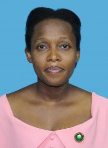
Josiane UWINEZA
Research Engineer
Python, Data science, Machine Learning, Deep Learning, Computer Vision, Prayer
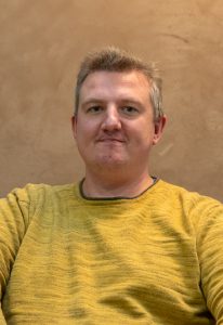
Didier WECKMANN
Senior Software Developer
C++, Python, JavaScript, Software Architecture, Continuous Integration, Science Fanboy
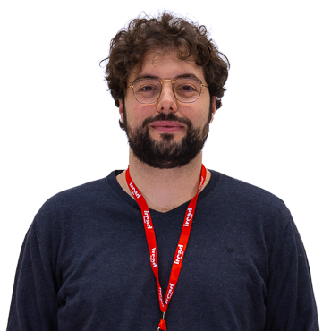
Flavio MILANA
Fellow
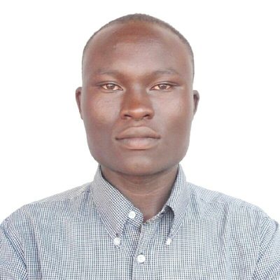
Cyriaque ZIRIMWABAGABO
Research Engineer
Yvonne KEEZA
Medical Imaging Annotator
Medical Image Data Analysis, Radiology, Ultrasound, Project Management, Image Protocol and Annotations, Medical Technology Enthusiast, Salsa Dancer

Jean De Dieu NIYONTEZE
Research Engineer
Grace UFITINEMA
Medical Imaging Annotator
Medical Image Analysis, Radiography, Ultrasound, Annotation, Basketball-Lover

Luis MENDOZA
Senior Software Developer
Qt, C++, Computer Vision, Another guitar-playing, Football-loving latino

Baptiste PODVIN
Phd. Student
Shamim SEDGHI
Master Student
Nicolas PAPIER
Senior Software Developer
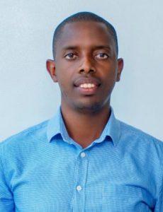
Florien UJEMURWEGO
Medical Imaging Annotator
Medical Image Analysis, Radiography, Ultrasound, Annotation, Medical Image Management, Swimming-Love
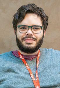
Güinther SAIBRO
Research Engineer
Python, Deep Learning, Statistics, Medical Image Analysis, Ultrasound, Cycling
















Don’t miss the latest news from IRCAD.
Sign up here for our newsletters and communication mailings
to stay informed about us and our courses
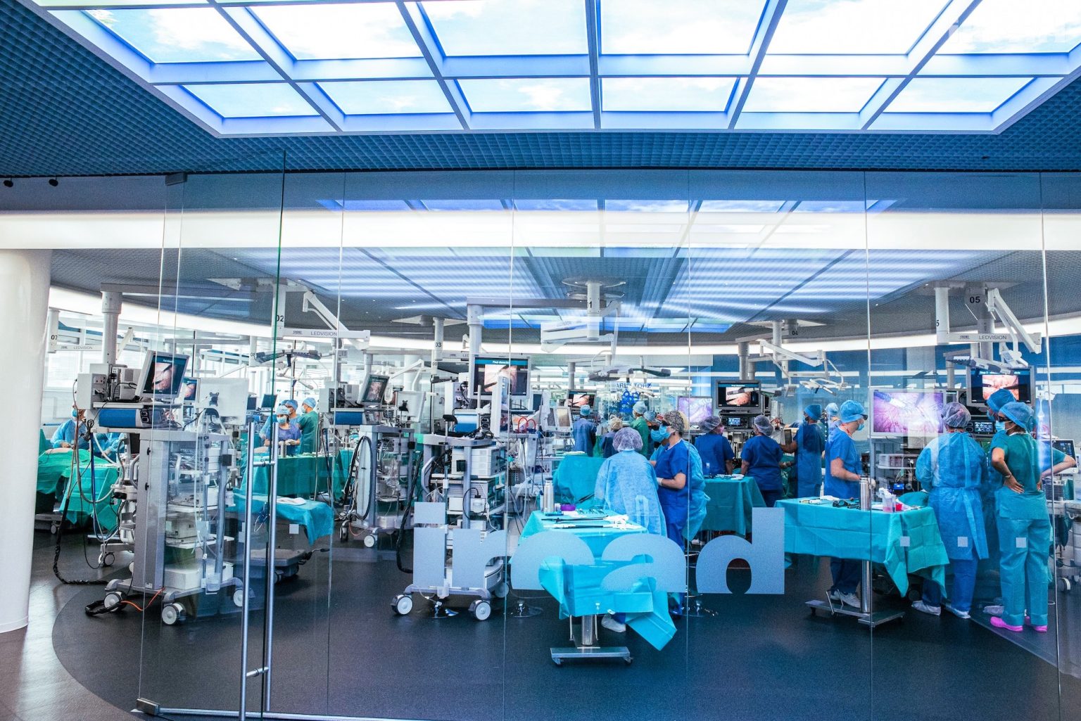
Please note that the IRCAD administrative board and staff are closely monitoring the evolving COVID-19 situation, in full compliance with all applicable laws and regulations in France. The health, safety, and well-being of our participants, experts and staff are our top priority!
Despite the current context, the IRCAD stands firmly by your side to help you acquire knowledge and skills. Come and join us !
We would like to draw your attention that the « Vaccine Pass » is now mandatory in France since end of January 2022 and replaces the former « Health Pass » to access places that are open to the public, such as cinemas, museums, cafés and restaurants, hotels as well as the IRCAD Institute which welcomes participants in the framework of its courses and seminars. Thus, a PCR test without vaccination is no longer sufficient to take part in our courses.
The vaccine pass includes a proof of the following (one of the 3 items is sufficient):
Further information about the new vaccine pass can be found at :
We very much hope to be able to count on your kind understanding of those rules which have been set by the French Government and which our Institute is required to apply