Building on the momentum of previous years, IRCAD enters 2025 with dynamic prospects, both nationally and internationally. Following the opening of IRCAD India in 2024, two new institutes will be inaugurated in China (Shanghai) and the United States (Charlotte), expanding the IRCAD network to a total of nine institutes.* The close collaboration within the network is a true driver of progress for patients worldwide, as illustrated by the Disrumpere research project, jointly conducted by IRCAD France and IRCAD Africa (Rwanda). This project aims to improve ultrasound access for African patients and, in the long term, for patients in medical deserts. We will soon be sharing updates on this project.
In 2025, thanks to new industrial partnerships, we will further enhance our research and training capacities with the creation of new robotic platforms that are increasingly innovative and diverse in their applications. IRCAD is now the world’s largest surgical robotics training platform, and we will have the honor of hosting the Surgical Robotics Summit (SRS) in Strasbourg this July.
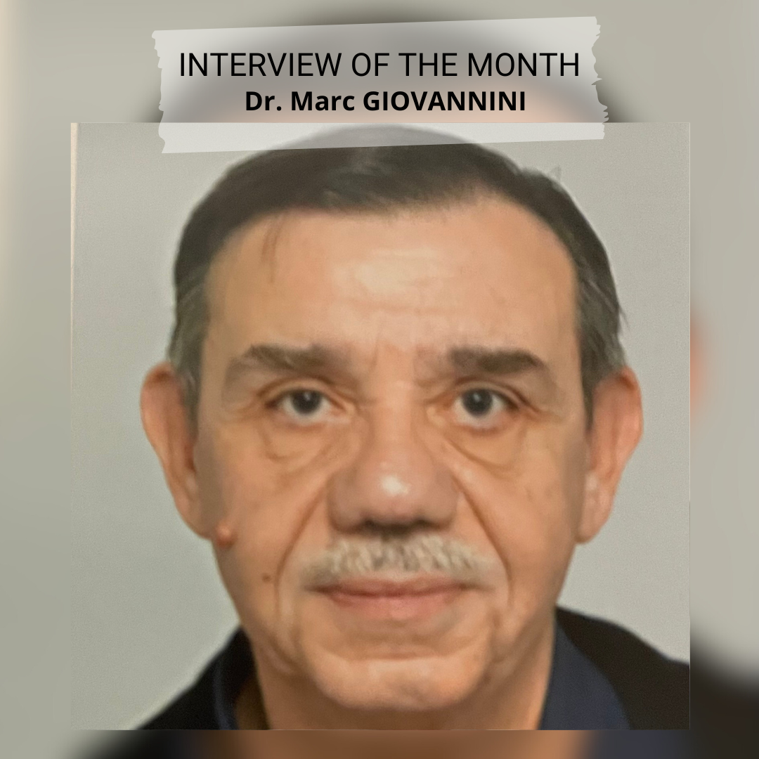
This year will also mark the expansion of our activities in the field of digestive endoscopy, with the launch of an annual training program covering both diagnostic and therapeutic aspects, as well as research initiatives incorporating artificial intelligence and augmented reality. To oversee these activities, Dr. Marc Giovannini has recently joined IRCAD France, bringing extensive expertise to the field. Notably, he previously served as the Head of the Digestive Endoscopy Department at the Paoli-Calmettes Institute in Marseille. I am delighted to give him the floor in this first newsletter of 2025 to introduce his priorities and projects.
*France, Taiwan, Brazil (2), Lebanon, Africa, India, China, United States

Professor Jacques Marescaux
President & Founder of IRCAD
Digestive endoscopy at IRCAD: new advances in research & training
Dr Marc GIOVANNINI
Former Head of the Digestive Endoscopy Department at the Paoli-Calmettes Institute (Marseille) & Co-Director of IRCAD’s Digestive Endoscopy Courses
Dr Marc Giovannini: IRCAD is a pioneer in minimally invasive and robotic surgery, having gained unique expertise in setting up training programs over the years. Since digestive endoscopy requires similarly demanding learning processes, the goal is to apply the same structured approach.
Previously, a few courses at IRCAD covered digestive endoscopy, but these were sporadic and focused on specific topics such as superficial tumor resection. My mission is to establish a structured and sustainable training program, particularly in therapeutic endoscopy, which is rapidly growing due to its many benefits for patients.
For 2025, four training sessions are already planned, ranging from basic skills—primarily for residents, covering endoscope handling, image recognition, and performing biopsies—to advanced courses on topics like echoendoscopy.
Moreover, IRCAD’s network of mirror institutes worldwide will enable us to export the courses we develop to other regions, particularly Africa, where access to digestive endoscopy remains limited. In the coming months, we will conduct the first digestive endoscopy course at IRCAD Africa (Rwanda) and are also in discussions with IRCAD Taiwan to introduce training there. The global demand for training in gastroenterology and flexible endoscopy is substantial, and contributing to meeting this demand is particularly motivating for me.
Additionally, IRCAD has a strong high-level research program, and Professor Marescaux invited me to work on projects combining endoscopy with artificial intelligence and robotics, which I find particularly exciting.
I also have a personal priority: the development of non-animal, reusable learning models that replicate the texture of human organs. Our first focus will be biliary drainage procedures under echoendoscopy, which currently lack a realistic training model—even animal models are unsatisfactory. Creating reusable models that allow specialists to repeat procedures until they achieve mastery would be a revolutionary breakthrough. These models could even extend beyond digestive endoscopy to benefit other medical fields.
Pr. M. G. : In most countries, both digestive endoscopy and digestive surgery are performed by the same specialist. For example, in Brazil, patient management is organized by organ, meaning that a colon specialist handles colonoscopy screenings, polyp resections, and surgical procedures for advanced cancers. In some Nordic countries, surgeons themselves perform endoscopy.
In France, however, there is a clear separation between digestive surgeons and gastroenterologists specialized in endoscopy, mainly due to their training paths. French digestive surgeons do not undergo dedicated endoscopy training, while gastroenterologists learn endoscopic techniques but do not train in surgery. Despite this separation, collaboration occurs when necessary.
This variation in international practices does not impact our training programs. IRCAD’s curriculum is designed to be universal, accommodating specialists from around the world.
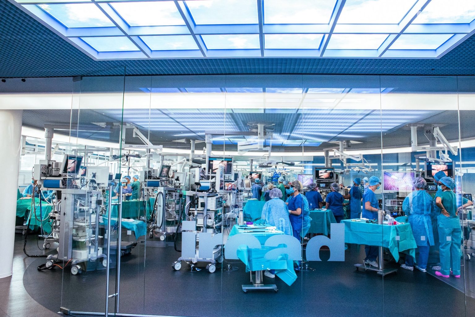
IRCAD offers a state-of-the-art digestive endoscopy facility, featuring 20 endoscopy towers and 3 echoendoscopy towers, along with other cutting-edge equipment. This unique training environment enables specialists to master both basic and advanced techniques while also training on different brands of endoscopic equipment.
Echoendoscopy combines endoscopy with ultrasound, allowing for a detailed ultrasonic analysis of the digestive wall and nearby organs. It is the most effective method for detecting tumors in the digestive tract (esophagus, stomach, duodenum) and surrounding organs (gallbladder, bile ducts, pancreas, liver, spleen, kidneys). It also facilitates biopsies by guiding a needle inside the endoscope.
Initially, echoendoscopy was purely a diagnostic tool, but technological advances have enabled its therapeutic applications. It now plays a crucial role in pancreatic cyst and abscess drainage, bile and pancreatic duct obstruction clearance, and targeted tumor destruction using radiofrequency ablation.
Training in echoendoscopy is highly demanding, requiring multiple attempts to achieve proficiency. It is also essential to train nurses, who play a key role in assisting with the procedure.
At IRCAD, we have developed a comprehensive training program covering the fundamentals of echoendoscopy, hands-on procedures, and therapeutic applications. Proper training is crucial because biopsies directly impact treatment decisions. For example, in pancreatic cancer, studies have shown that biopsies performed under CT scan increase the risk of cancer cell dissemination, as the needle passes through multiple organs. In contrast, echoendoscopy minimizes this risk by navigating through natural pathways.
This safe and precise technique represents a significant advancement—but only if practitioners receive proper training, which remains IRCAD’s top priority.
Research and development are at the heart of IRCAD, which remains open to all innovations with the ability to develop and evaluate them.
Several projects focus on augmented reality to further improve navigation inside the human body, particularly in echoendoscopy. The goal is to merge echoendoscopic images with images from other examinations. For example, if a patient has already undergone a CT scan that identifies the lesion to be biopsied, a navigation system that fuses real-time echoendoscopy images with those from the scan allows for even greater precision in targeting. We are working with multiple companies to develop and assess these advanced image fusion navigation systems.
Other projects focus on the use of artificial intelligence for both diagnostic and therapeutic purposes. In endoscopy, AI is already regularly used to interpret polyp images, as we have a sufficient dataset. In echoendoscopy, AI must assist in diagnosing small or ambiguous lesions and highlight areas that are difficult to see. Combining different types of imaging appears promising, and we are actively working on this.
Another important aspect is using artificial intelligence to ensure that an examination is complete and that the gastroenterologist has visualized all necessary areas. In echoendoscopy, for instance, a system can activate indicators as the pancreas is progressively examined (hook, head, body, and tail), confirming that the entire organ has been reviewed. Other systems can provide a percentage of the organ visualized, whether it be the colon or stomach. Developing this kind of exhaustive visualization is essential both for training and for patient care, ensuring that the examination has been thorough. If an organ has not been fully visualized and no further improvement is possible, knowing this allows for adjustments in patient management accordingly.
Artificial intelligence can also assist beginners in echoendoscopy by helping them confirm that they are correctly viewing the organ or pinpointing their exact location if they feel uncertain. For example, the system can outline the pancreas in yellow for better identification.
We are also working on projects related to robotic assistance, which requires modifications to the endoscope, as current devices lack triangulation and 3D vision. Robotic development will largely depend on the ability to miniaturize equipment, as space is very limited in the anatomical areas where we operate. In the rectum and stomach, slightly larger equipment could be used, with small robotic arms deployed for dissections. However, in the esophagus, duodenum, and even the colon, space constraints make this challenging today, but technological advancements may make it feasible in the future.
In all our projects, we are particularly focused on assessing the potential benefit for the patient. It is not uncommon for companies to present us with technologically innovative developments that, in our view, do not offer real improvements in patient care. It is crucial not to be swayed by the appeal of novelty but to always prioritize the patient’s best interests. We must constantly ask ourselves what will truly provide the best service to the patient – this is the core of our mission.
About IRCAD:
Founded in 1994 by Professor Jacques Marescaux, IRCAD is an internationally renowned institute dedicated to training and research in minimally invasive surgery. Each year, 8,800 surgeons from around the world train at the Strasbourg institute, either in-person or via WebSurg, a free online university with over 470,000 registered members worldwide.
For more information, visit https://www.ircad.fr/en/
We hope you enjoyed this 17th edition of the IRCAD newsletter.
For any inquiries, subscriptions, or unsubscriptions, please contact:
FINN Partners – sante@finnpartners.com